The Cardiovascular System

at a Glance

Fourth EditionPhilip I. Aaronson, Jeremy P.T. Ward & Michelle J. Connelly

Case Studies

Case 2 – Valvular heart disease

Show Image

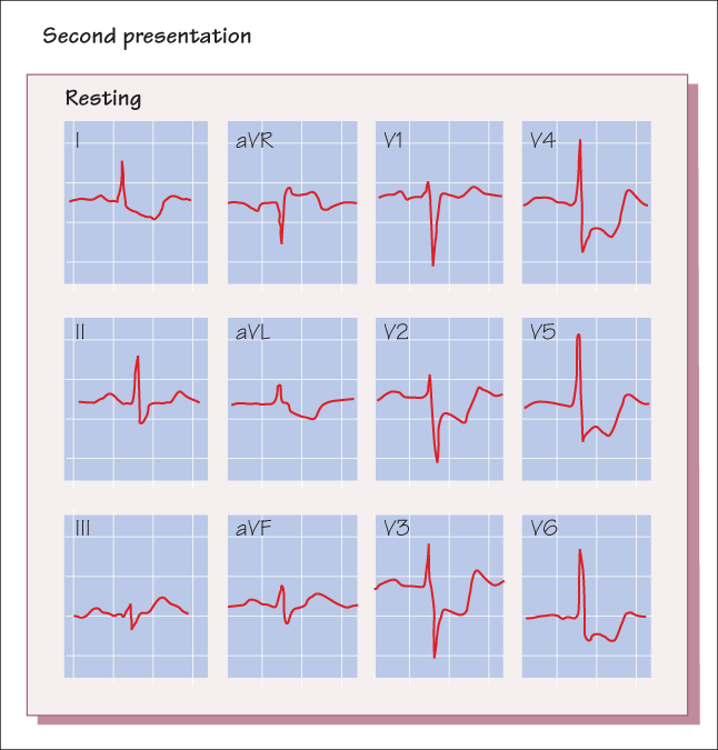
You are asked to supervise an exercise stress test on a 65-year-old man. He saw his doctor last week for exertional chest pain and mild dyspnoea. He has had chest discomfort for about a year, but the increased frequency of angina prompted him to see his doctor. He has chest pain when he walks more than one block, and if he continues he becomes breathless. He never has chest pain or dyspnoea at rest. He has no ankle swelling, orthopnoea or paroxysmal nocturnal dyspnoea. When you examine him before the stress test, his blood pressure is 120/86 mmHg heart rate 82 and regular, jugular venous pressure 5 cmH2O and lungs are clear. His apex beat is slightly lateral to the midclavicular line and mildly sustained. He has a normal S1 and a single S2. An S4 gallop is noted. He has a soft crescendo–decrescendo systolic murmur, best heard at the upper right sternal border, radiating to the carotids and the apex. The carotid pulses are delayed and diminished.

You call the referring doctor to discuss the signs and symptoms, cancel the stress test and perform an echocardiogram instead.

  • (a) What is the likely diagnosis based on the physical examination? What are the pathophysiological mechanisms underlying these findings?

    He is likely to have severe aortic stenosis. The crescendo–decrescendo systolic murmur usually arises from stenosis of either the aortic or pulmonary valve. The stenotic valve creates turbulence during ejection that causes a murmur. The murmur gets louder as flow increases during ejection, then diminishes as flow decreases. This murmur is transmitted to the carotid arteries because of the high velocity of the ejected blood. The apical murmur is probably caused by high frequency vibration of the aortic valve during ejection and can sometimes be louder than that at the sternal border.

    Aortic stenosis causes left ventricular hypertrophy, which displaces the apex beat laterally. The apex beat can be sustained because the ventricle takes longer to empty. The carotid pulse contour reflects flow across the aortic valve. Because peak ejection flow is delayed and decreased in aortic stenosis, the carotid pulses are delayed and diminished.

  • (b) He only had a soft systolic murmur. If you knew that his murmur last year was louder and harsher in intensity, would this have reassured you? What could go wrong if he did do the stress test?

    The loudness of murmur depends on flow across the valve; therefore it may not reflect the severity of valve stenosis. In the case of aortic stenosis, other physical examination parameters are more indicative of the severity of valve stenosis. As valve area becomes smaller, the peak loudness of the murmur occurs later in systole. This is presumably due to the difficulty in opening the valve. In severe aortic stenosis left ventricular stroke volume is decreased. This manifests as low-amplitude carotid pulsation with a delayed peak (pulsus parvus et tardus).

    Normally, the aortic valve closes before the pulmonary valve, causing splitting of S2. During inspiration more blood volume is returned to the right heart and the pulmonary valve closes even later. The increased splitting of S2 during inspiration is termed physiological splitting. In severe aortic stenosis, the ejection time becomes longer and the aortic valve closes later. This may eliminate splitting of S2 (single S2), or create paradoxical splitting.

    If this murmur was previously louder, it would suggest that flow across the valve has decreased as the stenosis worsened. Decrease in flow (lower cardiac output) is an ominous sign of a failing left ventricle or a decrease in valve size. This is not reassuring.

    During exercise, blood vessels dilate and peripheral resistance decreases. When the peripheral resistance falls, the blood pressure tends to fall. Normal individuals compensate by increasing stroke volume and heart rate. However, this patient cannot increase his stroke volume because of the tight aortic valve. When blood pressure falls, coronary perfusion decreases and the subendocardial area of the hypertrophied ventricle is not perfused adequately. As heart rate and oxygen demand increase, the supply and demand mismatch may worsen and cause ischaemia. If a significant portion of the myocardium is affected, cardiac output can fall, causing a further drop in coronary perfusion and further worsening ischaemia. This vicious cycle can continue until the patient drops dead, an event that may not look particularly good on your record.

  • (c) What was likely to be observed on the echocardiogram and why?

    The echocardiogram showed uniform left ventricular hypertrophy. His left ventricular ejection fraction was estimated at 45%, and the aortic valve appeared calcified with impaired cusp mobility. Doppler studies showed the peak instantaneous gradient across the aortic valve was 98 mmHg with a mean gradient of 68 mmHg. Estimated aortic valve area was 0.6 cm2.

    The most common cause of aortic stenosis in this age group is calcific degeneration of the aortic cusps. The valvular pathology leads to left ventricular hypertrophy, a pressure gradient and impaired emptying of the chamber, which were all demonstrated on the echocardiogram.

  • (d) Catheterization data The patient is formally referred to you, and you recommend valve replacement. He undergoes cardiac catheterization, and the haemodynamic data are shown in Figure Case 2. Cardiac output is 5.2 L/min and heart rate is 77. There is no significant coronary artery disease.

    The aortic valve area can be estimated by the simplified formula:
    Valve area = cardiac output/

    Based on the data given, what is his estimated valve area?

    Using the formula, the estimated valve area is 0.62 cm2. This formula is a simplified version of the Gorlin formula, which states that the valve area is related to cardiac output, and inversely related to mean pressure gradient, systolic ejection period and the gravitational acceleration constant. In most cases, the systolic ejection period multiplied by the acceleration constant is close to 1, so that term is not used in the estimate formula.

  • (e) When admitted for surgery, he complains of chest pain. The intern orders sublingual nitroglycerin for him. Why is this a bad idea? What is the chest pain due to? What therapeutic options are available for protracted chest pain in this case?

    This patient’s left ventricle (LV) needs all the help it can get to eject an adequate supply of blood through the pinhole-sized aortic valve. Nitroglycerin is primarily a venodilator, and it will pool his blood in the veins. This will reduce blood returning to the heart and decrease preload. Even though the ejection fraction is only 45%, his thick LV still has high contractility and a steep end-systolic pressure–volume relation. This means that he is very sensitive to preload changes, and a slight drop in preload volume can drop blood pressure significantly, which will decrease coronary perfusion, worsen subendocardial ischaemia, decrease systolic dysfunction, lower blood pressure, and so on. Thus, nitrates are a bad idea. Beta-adrenergic blockers or calcium-channel blockers are also not good because they can reduce contractility. Calcium-channel blockers can also dilate peripheral arterioles and reduce blood pressure further.

    What can you do? You may actually give some fluid to increase preload and increase cardiac output. The other mode of therapy, if the valve cannot be replaced promptly, is intra-aortic balloon counterpulsation. This can improve coronary perfusion in some cases and relieve subendocardial ischaemia.

    Concluding remarks
    Valve replacement is best performed before development of ventricular dysfunction or pulmonary hypertension. Either tissue (xenografts, commonly from pig) or mechanical valves can be used. Although the latter are harder wearing, they can promote thrombosis, so anticoagulant therapy will be required.

Print Answers | « Previous Case | Next Case »

twitter