-
(a) Is this angina pectoris?
Show Answer
This sounds like angina. The location, radiation and type of discomfort are consistent with cardiac ischaemic pain. However, the traditional dividing line between ‘angina’ and possible MI is 30 min of discomfort. Because her discomfort lasted more than 30 min, it is possible she suffered an MI, even with a normal ECG. To determine whether an MI occurred, look for evidence of myocardial damage such as cardiac-specific serum enzymes.
-
(b) What predisposing factors does she have for coronary artery disease (CAD)? What other risk factors might she have? Could this have been an MI?
Show Answer
Her risk factors for CAD are smoking and being postmenopausal. Her family history is not positive for premature CAD, because her father was 74 when he had his MI. She does not have hypertension. We do not yet know her cholesterol levels, or whether she is diabetic.
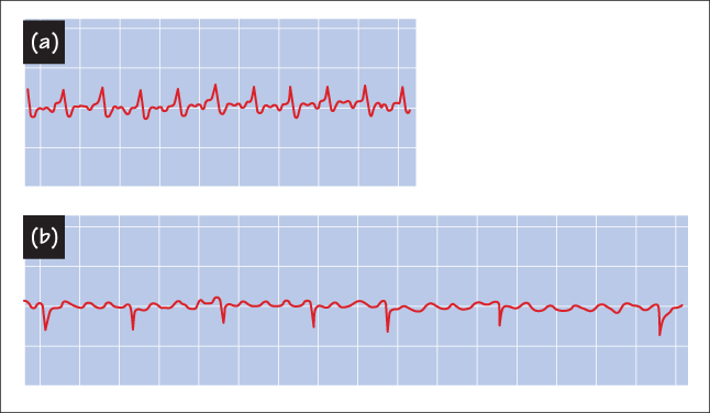
- She is admitted for monitoring. Serial serum enzyme tests are negative, and ECG remains normal after 18 h. Fasting serum glucose is 92, and fasting lipid profile is: total cholesterol 198, high-density lipoprotein (HDL) 36, low-density lipoprotein (LDL) 137 and triglycerides 126. She undergoes a graded exercise test for 12 min, and has chest discomfort with the ECG abnormalities shown in Figure Case 3 (b). Her blood pressure rose from 124/78 to 180/76 mmHg at peak exercise. Her heart rate rose from 78 to 143. The chest discomfort resolved within 5 min of stopping exercise, and the ECG returned to normal. Coronary angiography showed a focal 70% narrowing in the right coronary artery and a focal 30% narrowing in the left anterior descending artery (LAD). She is treated with anti-ischaemic medicines, advised on low-cholesterol diets and aerobic exercise, instructed on the use of sublingual nitroglycerin, started on aspirin and atenolol, and discharged. She agrees to stop smoking. She will return for follow-up in 7 days for another exercise test.
-
(c) What serum enzymes are useful in diagnosing myocardial damage?
Show Answer
Increased serum levels of the cardiac (MB) isoform of creatine kinase and of the cardiac troponins T and I are indicative of myocardial damage. Unlike CK-MB, troponins are selective for cardiac injury, and are therefore now the preferred markers.
-
(d) Why not perform angioplasty on the coronary lesions?
Show Answer
She does not have unstable angina at this point, and her effort tolerance is reasonable. Such patients have a good prognosis whether they take medicines or have single-vessel angioplasty. A recent study showed that angioplasty was no better than medicines for single-vessel CAD.
-
(e) Her cholesterol is not high. Why a low-cholesterol diet? Would lowering blood cholesterol help her existing CAD? What other risk factors can she modify?
Show Answer
Her cholesterol was not high, but studies indicate that lowering LDL cholesterol below ~110 mg/dL may stabilize coronary lesions and possibly cause regression of CAD. However, greater effects were seen on reduced incidence of unstable angina or MI. These may be due to reducing the lipid-rich area of plaques and making them less susceptible to rupture.
The most important thing is to stop smoking. Reducing cholesterol is a secondary prevention. Another risk factor is lack of oestrogen. Postmenopausal women with CAD may benefit from hormone replacement therapy.
-
(f) Why perform a follow-up exercise test?
Show Answer
Reasons for exercise testing are diagnosis, prognosis and to test effectiveness of treatment. Here, we want to know if her anti-anginal regimen is adequate.
- Second presentation : The patient does well on the medicines and stops smoking. She has no chest discomfort, even during vigorous exercise. However, within the week she is awakened from sleep by the same chest discomfort. As nitroglycerin is ineffective, she is brought to hospital. Her ECG is shown in Figure Case 3 (c). She is given oxygen, aspirin, intravenous nitroglycerin and heparin, and her chest discomfort then recedes. It lasted for 35 min. Because of recurrent chest discomfort in hospital, she has another coronary angiogram. This shows the same 70% narrowing in the right coronary artery, but the 30% LAD narrowing has now grown to 99%. Cardiac enzymes are still negative.
-
(g) What does the new ECG show? What is your diagnosis? How is the pathophysiology of the current chest discomfort different from her previous effort-related angina?
Show Answer
The ECG shows marked ST segment depression in the precordial (V1–V6) and lateral (I and aVL) leads. She has cardiac ischaemia, due to either unstable angina or an MI. The chest discomfort occurred during sleep, indicating that it is unstable rather then stable angina. She is unlikely to have had an MI, as her cardiac enzymes are not elevated.
Cardiac ischaemia occurs when myocardial oxygen demand exceeds supply. This typically happens with exertion in CAD patients, because blood flow through the stenosis is usually adequate during rest. Rest ischaemia is caused when the oxygen supply falls below that required for even basal myocardial metabolic needs. This implies an occlusion more serious than that caused simply by a structural stenosis. It is believed that unstable angina is initiated by plaque rupture, causing aggregation of platelets and fibrin deposition. This can cause direct occlusion of the artery, and also trigger vasospasm.
-
(h) Do the ECG abnormalities correlate with the coronary angiogram? What is the ‘culprit’ lesion? How could the LAD lesion progress so fast? Why use aspirin and heparin? How about thrombolytic agents?
Show Answer
The angiogram suggests the ‘culprit’ is the LAD lesion. It is serious enough to cause rest angina. The ECG abnormalities are consistent with the ischaemic area being that served by the LAD. Unstable angina involves platelets and the clotting cascade. Thus, it makes sense to use inhibitors of platelet aggregation such as aspirin and clopidogrel. Clot formation and dissolution are dynamic processes, and although antiplatelet drugs cannot directly disperse clots, they can promote resolution. Thrombolytics make theoretical sense, but are not used because in trials no improvement in outcome of unstable angina patients was seen, and morbidity was higher.