-
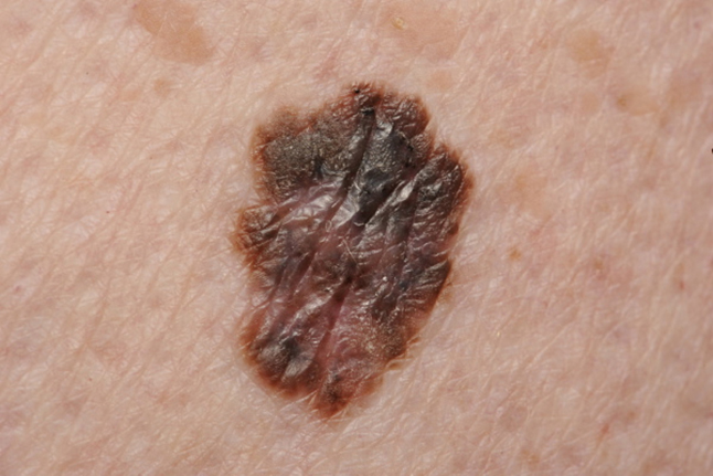
(a) What is the diagnosis?
Show Answer
This is a malignant melanoma. Other pigmented lesions that most commonly cause confusion with malignant melanoma include moles, pigmented basal cell carcinomas and seborrhoeic keratoses. Malignant melanoma (MM) arises from epidermal melanocytes. These tumours may arise within long-standing or new pigmented lesions.
-
(b) What features would suggest this diagnosis?
Show Answer
Any mole that stands out as being irregular compared with other moles present should always be treated with a high degree of suspicion. Any change in size, shape, colour with associated redness, itching or bleeding should be enquired about.
-
(c) How will you treat this lady?
Show Answer
The clinical diagnosis of suspected melanoma needs to be urgently confirmed with histology. This woman’s mole should be removed fully with primary excision with a 2 mm margin of normal surrounding skin followed by wide local excision, which reduces local recurrence.
-
(d) What does the prognosis depend on?
Show Answer
The Breslow thickness measures the distance of the deepest invasive area of the primary tumour (in millimetres) from the epidermal granular layer. Lesions < 1 mm thick are considered lower risk and > 4 mm are higher risk. Five-year survival falls with increased thickness of the tumour.
-
(e) What monitoring does she need?
Show Answer
Patients should be taught self-examination as early detection of recurrences is important. Patients with invasive MM should be followed up 3-monthly for 3 years and discharged if < 1 mm thickness. Thicker lesions > 1 mm should be followed up for further 2 years at 6-monthly intervals.