- Home
- Cases
- Revision notes
- Your feedback
- Become a reviewer
- 'At a Glance' series
- More student books
- Student Apps
- Join an e-mail list
You are 2 weeks into your new job as a foundation year house officer in geriatric medicine. An 84-year-old woman has been transferred to your hospital for rehabilitation following a successful tissue aortic valve re-placement at a tertiary centre. On examination she is comfortable at rest, her hands are cool and her JVP is elevated to her jaw. Corneal arcus is present. Inspection of her precordium reveals a fresh midline sternotomy scar that is healing well. Her apex beat is located in the sixth intercostal space, mid-axillary line. Auscultation of her precordium reveals normal heart sounds. Her chest is notable for stony dullness at the left base and bibasal crepitations. She has pitting oedema to her thighs. ECG shows rate-controlled atrial fibrillation and her chest X-ray shows cardiomegaly, bilateral bat’s wing shadowing and bilateral pleural effusions (Case 5 figure). On the ward round, she complains of a poor night’s sleep and being ‘unable to catch my breath’. A recent echocardiogram showed an ejection fraction of 35%.
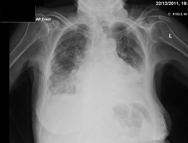
AP erect film. Bilateral pleural effusions with adjacent lung atelectasis and generous cardiac silhouette. Sternotomy wires in situ. There is also separation of the left acromioclavicular joint.
1. What is the JVP a measure of?
2. Which vessel are we looking at when we assess the JVP?
3. What is corneal arcus?
4 .Is her apex beat displaced?
5. What is the diagnosis in this woman?
6. What do cardiomegaly, bibasal shadowing and the pleural effusion on her chest X-ray represent?
7. How do you manage this patient?
8. Do all patients with cardiac failure have a reduced ejection fraction on echocardiogram?
See Chapters 46 and 47.