-
(a) What does the III/IV holosystolic murmur indicate?
Show Answer
This indicates a moderate murmur of mitral regurgitation (see Chapters 30 and 54).
-
(b) Is this tachycardia more likely supraventricular or ventricular? Why? What is the axis and the QRS morphology?
Show Answer
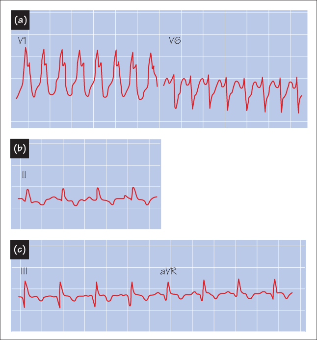
This rhythm is most likely ventricular in origin. When a patient has a wide-complex tachycardia, the differential diagnosis is ventricular tachycardia (VT) vs supraventricular tachycardia with aberrant conduction (i.e. the tachycardia is conducted with a bundle branch block). A wide variety of clinical and ECG criteria aid in distinguishing between these possibilities. The most important clinical criterion is whether the patient has a history of heart disease (see below). If this is the case, and in particular if there is a history of MI, the most likely origin of the tachycardia is ventricular, because an infarct creates a substrate for re-entry. ECG criteria that suggest a ventricular origin include: (i) a very wide QRS complex; (ii) an extreme axis; (iii) evidence of atrial–ventricular dissociation; (iv) certain specific QRS morphologies. This patient’s QRS morphology is that of a right bundle. Note the terminal S waves in V6, and the R in V1. The extreme axis, the very wide QRS duration and the morphology (R/S < 1 in V6 and the large primary R in V1) suggest the rhythm is VT.
-
(c) Why is he short of breath and hypotensive? Why is his JVP elevated?
Show Answer
He is short of breath and hypotensive because the tachycardia does not allow enough time for ventricular filling. Therefore, cardiac output is low, causing hypotension, and left atrial pressure is high. The high left atrial pressure has backed up into his pulmonary capillaries, causing pulmonary oedema and breathlessness. His increased JVP is caused by raised central venous pressure, indicative of inadequate right ventricular function. His arrhythmia is therefore causing congestive heart failure.
-
(d) What would be appropriate treatment?
Show Answer
Treatment requires cardioversion to sinus rhythm with either appropriate drugs (lidocaine, procainamide or amiodarone) or electrical cardioversion. Because this patient’s symptoms indicate that his arrhythmia is causing his heart to fail, immediate conversion to sinus rhythm is mandatory. Therefore in this case electrical cardioversion is preferable.
- Results of treatment The patient receives lidocaine. This fails to convert him to sinus rhythm and he is therefore electrically cardioverted. The resulting ECG is shown in Figure Case 5 (b) and (c).
-
(e) What is the rhythm now?
Show Answer
The rhythm is now sinus, with the rate of 95 beats/min.
-
(f) What is the likely cause of his acute presentation?
Show Answer
He has evidence on his ECG of an inferior wall MI (note the Q waves in leads II, III, aVF with slight ST elevation). The aetiology of his ventricular tachycardia is likely to be re-entry into left ventricular (LV) scar caused by an undetected recent acute MI.
-
(g) What would be the appropriate evaluation with this information?
Show Answer
Following stabilization, appropriate treatment includes evaluation of LV function and of coronary anatomy.
Concluding remarks - In this case, the patient had severe inoperable coronary artery disease and poor LV function. Because a poor ejection fraction identifies high risk for recurrent VT and sudden death, the patient underwent electrophysiology testing and was readily inducible into hypotensive VT. This indicated: (i) that he was at a high risk of spontaneously developing VT again; and (ii) that his tachycardia was fast enough to lower his cardiac output dangerously. His already poor LV function meant that he would not be able to tolerate tachycardia. He therefore subsequently underwent successful placement of an internal cardioverter defibrillator and has since been stable.


